Low back pain

WORK IN PROGRESS
WORKS IN PROGRESS.... ITEM IN PROCESSING...

NOTES ON THE STRUCTURE OF THE RACHIS

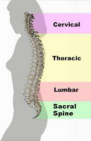
The vertebral column, also called rachis, is composed of 33-34 vertebrae. On the basis of the different configuration of the vertebrae it can be divided into four sections which, named in the cranio-caudal sense are: cervical, dorsal, lumbar, sacred. The first and third under physiological conditions form lordotic curves (curves of the column to posterior concavity) , while the other two form kyphotic curves (curves of the column with anterior concavity).

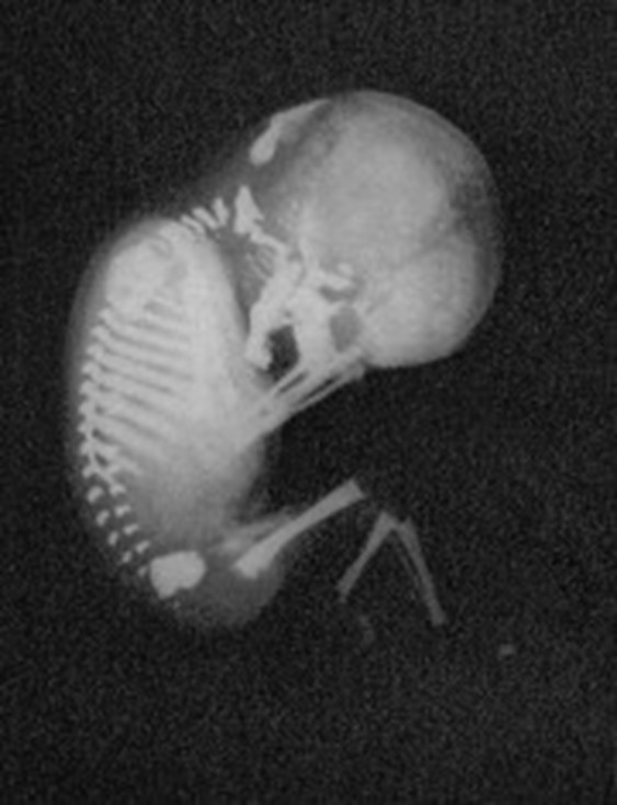
In the fetus, the spine appears as a single C-shaped curve. Even before birth, a cervical countercurve begins to appear (cervical lordosis), which will then develop after birth in parallel with the stages of acquisition of the ability to extend the head[1]Bagnall, K. M., P. F. Harris, and P. R. Jones. “A radiographic study of the human fetal spine. 1. The development of the secondary cervical curvature.” Journal of anatomy 123.Pt 3 1977: … Continue reading [2]Been, Ella, Sara Shefi, and Michalle Soudack. “Cervical lordosis: the effect of age and gender.” The Spine Journal 17.6 2017: 880-888.. At the lumbar countercurve (lumbar lordosis) it develops in relation to the achievement of the upright position and walking.

The lordoses (cervical and lumbar), also called secondary curves, they allow to recover the horizontality of the gaze by counterbalancing the inclination given by the kyphotic curves. (dorsal and sacral), also called primary curves due to the fact which preserve the original trend of the first large fetal C, among other things they have the task of welcoming and protecting in their heart concavity, lungs and pelvic organs. The alternation of the primary and secondary curves gives the structure of the spine the ability to cushion the weight and to bear considerable levels of load stress.

The four curves of the spine: an alternation of primary and secondary curves
Bagnall, K. M., P. F. Harris, and P. R. Jones. "A radiographic study of the human fetal spine. 1. The development of the secondary cervical curvature."

THE LUMBAR SPINE

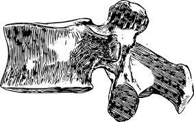
The lumbar spine is made up of 5 vertebrae (L1-L5). It articulates upstream with the dorsal rachis and downstream rests on the sacrum. The lumbar vertebrae have a robust body, adequate for their task of supporting the weight of a large part of the trunk, of the upper limbs and skull. This is the portion of the spine that bears the greatest load ever. The facet orientation of the lumbar vertebrae severely limits the possibility of rotational movement at this level to 1° per vertebra, for a total of 5° for the entire lumbar spine.

The spinal cord, which is that portion of the central nervous system that we can consider as the continuation of our brain outside the skull and which is contained within our spine, extends to the second lumbar vertebrae.

The vertebral column has two secondary curves, not present at birth: cervical and lumbar lordosis.
The lumbar vertebrae have some common characteristics: the large vertebral body and the orientation of the facet joints, arranged in a para-sagittal plane. This arrangement severely limits the rotational movements of this part of the spine.

LOMBALGIA

The pain syndromes affecting this section are subdivided, based on their topographic extent, in back pain, lombosciatalgie (of sciatica) e lombocruralgie (the cruralgie).

In low back pain the pain is limited to the lumbar region while in sciatica extends to the territory of the sciatic nerve and in the lombocruralgia to that of the crural nerve.

Low back pain is much more common than the other two syndromes[3]Morlachs, A. Mancini, C., and Antonio Mancini. “Orthopedic clinic” Piccin Publisher 1995.. It manifests itself with:

  • spontaneous pain of varying intensity localized in the lumbar area. It is accentuated by local pressure and attempts to mobilize the trunk[4]Morlachs, A. Mancini, C., and Antonio Mancini. “Orthopedic clinic” Piccin Publisher 1995..
  • contracture of the paravertebral muscles and position of the lumbar spine in slight forced anterior or lateral flexion[5]Morlachs, A. Mancini, C., and Antonio Mancini. “Orthopedic clinic” Piccin Publisher 1995.
  • pain aggravated by weight bearing mechanical, which increases in standing position, with minimal or no symptoms at rest[6]Carp, Year, et al. Management of degenerative disk disease and chronic low back pain. Orthopedic Clinics 42.4 2011 513-528. .
CLASSIFICATION OF LOWER BACK PAIN ACCORDING TO ETIOLOGY [7]Carp, Year, et al. Management of degenerative disk disease and chronic low back pain. Orthopedic Clinics 42.4 2011 513-528.
  • MECHANICS 80%-90% : degenerative disease of the intervertebral disc or facet joints, vertebral fracture, spinal deformity; I intervertebral discs they are the major tissue source of chronic low back pain (back pain lasting more than three months and/or recurring back pain): the most common finding that is highlighted by MRI in patients with chronic low back pain is the degeneration of one or more discs of the last three lumbar vertebrae.
  • NEUROGENA 5%-15% :. slipped disc, spinal canal stenosis;
  • NEOPLASMS, INFECTIONS, INFLAMMATORY SITUATIONS 1%-2%
  • REFERRED VISCERAL PAIN 1%-2%. Es.: gastrointestinal disease, kidney disease, abdominal aortic aneurysm;
  • OTHER 2%-4%. Es.: fibromyalgia, somatoform disorder;

From the causes of “mechanical low back pain” and of “lombalgia neurogena”.

  1. DEGENERATIVE DISC DISEASE
  2. FACET JOINT SYNDROME
  3. VERTEBRAL FRACTURE
  4. SPONDILOLISIS (VERTEBRAL FRACTURE) E SPONDILOLISTESI
  5. DEFORMED’ OF THE COLUMN: SCOLIOSIS
  6. SLIPPED DISC
  7. STENOSIS OF THE VERTEBRAL CANAL

1. MALATTIA YOUINTERVERTEBRAL DISC ENERATIVE

2. FACETS SYNDROME ARTICLES (FJS: Facet Joint Syndrome)[8]Brotzman, S. Brent, and Robert C. Manx. Clinical orthopaedic rehabilitation e-book. An evidence-based approach-expert consult. Elsevier Health Sciences, 2011. [9]Perolat, Romain, et al. Facet joint syndrome: from diagnosis to interventional management. Insights into imaging 9.5 2018 773-789..

The functional unit of the spine it's composed by 2 overlapping vertebrae that form a three-joint structure: an amphiarthrosis between the bodies of the vertebrae, which are articulated through the interposition of the vertebral disc, and two joints synovial (diarthrodia with capsular-ligament structures) between the posterior facet joints of the vertebrae. Degenerative changes in a joint affect the biomechanics of the entire complex[10]Perolat, Romain, et al. Facet joint syndrome: from diagnosis to interventional management. Insights into imaging 9.5 2018 773-789..

The orientation of the facet joints of the lumbar vertebrae on the one hand allows a greater range of motion of the lumbar spine in flexion and on the other hand contains its range in rotation. The vertebral facet joints function as a track for the movement of the spine and therefore have a fundamental role in the biomechanics of the spine. However, this function of theirs makes them able to be subjected to important forces and therefore be subject to the possibility of develop significant degenerative changes becoming a potential source of pain[11]Borenstein, David. Does osteoarthritis of the lumbar spine cause chronic low back pain?. Current pain and headache reports 8 2004 512-517.: capsule, synovium and subchondral bone are richly innervated by nociceptive fibers which are stimulated in case of degenerative and inflammatory processes but also in case of mechanical stretching.

SYMPTOMS

Unilateral or bilateral lumbar pain,. usually located in the lumbar region but in some cases it radiates to one or both buttocks, on the sides of the groin and thighs, up to above the knee[12]Manchikanti L, Singh V, Pampati V et al 2001 Evaluation of the relative contributions of various structures in chronic low back pain. Pain Physician 4 4 308–316. Iin some cases, FJS can mimic pain caused by herniated discs or root compression, with pain radiated beyond the knee[13]Brotzman, S. Brent, and Robert C. Manx. Clinical orthopaedic rehabilitation e-book. An evidence-based approach-expert consult. Elsevier Health Sciences, 2011. and the physical exam and medical history aren't enough to do the differential diagnosis. The symptomatology is typically aggravated by lumbar extension (which compresses increases the load on the facet joints). The onset of pain is acute when the’extension is combined with rotation. Lumbar flexion (which reduces the contact between the facet joints) relieves symptoms .

ETIOPATHOGENESIS

Among the main causes of FJS is the functional overload of the facet joints, which can be aggravated by factors such as obesity or the presence of inadequate muscles (sedentary people who perform little physical activity). L’osteoartrite (arthrosis) it is the most frequent form of pathology affecting the vertebral facet joints[14]Kalichman, Leonid, et al. Facet joint osteoarthritis and low back pain in the community-based population. Spine 33.23 2008 2560.. The arthrosis process in advanced stages can lead to the formation of synovial cysts.

Also rheumatoid arthritis and ankylosing spondylitis can lead to damage to the vertebral facet joints.

DEGENERATIVE OSTEOARTHRITIS OF THE FACETS JOINTS

Degenerative facet osteoarthritis (arthrosis) it is the most frequent cause of FJS and is closely related to disc degeneration intervertebral discs: ’80% about of patients with facet joint syndrome present obvious signs of a previous herniated disc[15]Brotzman, S. Brent, and Robert C. Manx. Clinical orthopaedic rehabilitation e-book. An evidence-based approach-expert consult. Elsevier Health Sciences, 2011.. The degenerative osteoarthritis process causes inflammation of the surrounding tissues that generates local pain[16]Perolat, Romain, et al. Facet joint syndrome: from diagnosis to interventional management. Insights into imaging 9.5 2018 773-789..

RISK FACTORS of DEGENERATIVE OSTEOARTHRITIS

  • the age
  • the sex
  • the spinal level (L4-L5)
  • the orientation of the facets (sagittally oriented)
  • presence of intervertebral disc degeneration. The latter factor is often related to the heavy work done before 20 years.

PURE SYNOVIALS[17]Perolat, Romain, et al. Facet joint syndrome: from diagnosis to interventional management. Insights into imaging 9.5 2018 773-789..

When the arthritic degenerative process of the facet joints is very advanced, it may happen that a herniation of the synovium occurs (synovial cyst) through the joint capsule. the synovial cysts typically cause radiculopathies due to compression of the nerve roots, particularly at the level of the lateral recesses. In some cases they can cause a symptomatic spinal stenosis.

DIAGNOSIS

The diagnosis of FJS is clinical but it is extremely difficult to differentiate it from other causes of low back pain. Furthermore, diagnostic imaging is not of much help because non there is a clear correlation between the clinical symptoms and the degenerative changes of the spine that are highlighted by the images[18]Manchikanti, Laxmaiah, et al. Management of lumbar zygapophysial facet joint pain. World journal of orthopedics 7.5 2016 315. [19]Kalichman L, Kim DH, That L, Germazi A, Hunter DJ 2010 Computed tomography-evaluated features of spinal degeneration. Prevalence, intercorrelation, and association with self-reported low back pain.… Continue reading. The painful response to maneuvers involving the mobilization of the vertebral facet joints, the positivity of the test in extension of the spine and the attenuation of the symptoms with flexion of the spine must in any case lead to the suspicion of FJS.

EPIDEMIOLOGY

It is estimated that the 15-40% of cases of chronic low back pain are caused by pathological processes affecting the facets vertebral joints.

TREATMENT

In case the rehabilitation treatments are not decisive, these patients may benefit from joint steroid injections [8] and/or specific interventions to eliminate pain in the facial joints, such as neurolysis [9].L

3. VERTEBRAL FRACTURE

4. SPONDILOLISIS (VERTEBRAL FRACTURE) E SPONDILOLISTESI

SPONDYLOLISTESIS

Spondylolisthesis is slippage of a vertebra in the sagittal plane, which in most cases is related to osteoarthritis of the facet joints. It occurs following a subluxation of the facet joints, related to an important and progressive loss of cartilage and joint remodeling, with segmental instability which causes a tension of the capsule. The level of the spine that is most affected is that of the L4-L5 segment, which turns out to be the same one that undergoes the major degenerative facet arthritic alterations. When spodylolisthesis occurs in young people it may be due to congenital anomalies, acute or stress fractures[20]Perolat, Romain, et al. Facet joint syndrome: from diagnosis to interventional management. Insights into imaging 9.5 2018 773-789..

In spondylolisthesis, the ROM of the lumbar spine is reduced, lat lumbar lordosis disappears (lumbosacral kyphosis) and a level prominence is highlighted sacral with a palpable "step".[21]Brotzman, S. Brent, and Robert C. Manx. Clinical orthopaedic rehabilitation e-book. An evidence-based approach-expert consult. Elsevier Health Sciences, 2011..

Back pain is present in the 75% of cases.

SPONDILOLISIS

Spondylolysis occurs most commonly in children little ones performing repeated push-ups and extensions of the column in the maximum degrees of movement (es., danza, artistic gymnastics,..). E’ was found a phereditary redeployment.

Symptoms usually include lower back pain and occasionally back pain in the buttocks and thighs without neurological deficits.

The "stork test" allows you to highlight pain from localized spondylolisthesis. The patient stands on one limb with the contralateral foot supported on the supporting knee. The patient then hyperextends the lumbar spine. A reproduction of lumbar pain of the patient indicates, until proven otherwise, a diagnosis of spondylolysis.

5. DEFORMED’ OF THE COLUMN: SCOLIOSIS

Directly consult the specific in-depth page for the topic by clicking WHO.

6. SLIPPED DISC

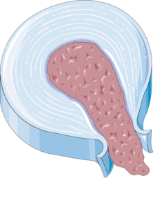
Beyond the 95% of the lumbar disc herniation happens at the level at L4-L5 (root L5) or at the level of L5-S1 (S1 root). The 75% of lumbar disc herniations resolve spontaneously within 6 months[22]Brotzman, S. Brent, and Robert C. Manx. Clinical orthopaedic rehabilitation e-book. An evidence-based approach-expert consult. Elsevier Health Sciences, 2011.. The herniated disc, depending on the anatomical relationships it establishes with the surrounding structures, can give rise to lumbar pain but also to sciatica. In this case the pain and paresthesias in the limbs are stronger than back pain. Acute disc herniation is usually characterized by the onset sudden sensation of lumbar pain and radicular pain to the lower limb[23]Brotzman, S. Brent, and Robert C. Manx. Clinical orthopaedic rehabilitation e-book. An evidence-based approach-expert consult. Elsevier Health Sciences, 2011.. The Valsalva test (ad is., sneeze, cough or defecation) leads to an exacerbation of pain.

(For further information click WHO for sciatica and WHO for disc herniation).

SLIPPED DISC. (Credits: Servier Laboratories; Source: https://smart.servier.com/category/anatomy-and-the-human-body/locomotor-system/bones/)

7. STENOSIS OF THE VERTEBRAL CANAL

Vertebral canal stenosis.

RISK FACTORS FOR LOWER BACK PAIN[24]Hoogendoorn, Wilhelmina E., et al. Systematic Review of Psychosocial Factors at Work and Private Life as Risk Factors for Back Pain. SPINE 25.16 2000 2114-2125. [25]Bernard BP, editor. Musculoskeletal disorders and workplace factors. A critical review of epidemiologic evidence for workrelated musculoskeletal disorders of the neck, upper extremity, and low back.… Continue reading [26]Burdorf A, Sorock G. Positive and negative evidence of risk factors for back disorders. Review. Scand J Work Environ Health 1997;23 4 243-56

  • age
  • sex: recent studies have shown that women tend to have episodes of longer duration and in a higher rate of worse outcomes than men. These results are part of a more general picture that shows that women, as a percentage, experience more intense pain, in a greater number of body districts, more frequently and for longer periods than men[27]Fulani, Wilco C., et al. “Influence of gender and other prognostic factors on outcome of sciatica.” Pain 138.1 2008: 180-191..
  • social class
  • performing activities that involve trunk twisting [28]Hoogendoorn, Wilhelmina E. Physical load during work and leisure time as risk factors for back pain. Scand J Work Environ Health 25.5 1999 387-403.
  • performing activities that involve heavy lifting[29]Hoogendoorn, Wilhelmina E. Physical load during work and leisure time as risk factors for back pain. Scand J Work Environ Health 25.5 1999 387-403.
  • performing activities that subject the body to vibrations: bus drivers, truckers…[30]Hoogendoorn, Wilhelmina E. Physical load during work and leisure time as risk factors for back pain. Scand J Work Environ Health 25.5 1999 387-403.
  • physically demanding professions[31]Hoogendoorn, Wilhelmina E. Physical load during work and leisure time as risk factors for back pain. Scand J Work Environ Health 25.5 1999 387-403.
  • low job satisfaction[32]Hoogendoorn, Wilhelmina E., et al. Systematic Review of Psychosocial Factors at Work and Private Life as Risk Factors for Back Pain. SPINE 25.16 2000 2114-2125.
  • jogging
  • Sedentary lifestyle
  • Obesity: both overweight and obesity appear to be risk factors for sciatica with a dose-response relationship (the risk increases with increasing weight). No difference in response was found between men and women[33]Plan, Rahman, et al. “Obesity as a risk factor for sciatica: a meta-analysis.” American journal of epidemiology 179.8 2014: 929-937..
  • Diabetes[34]Hassan, Ahmed, et al. Chronic low-back pain in adult with diabetes: NHANES 2009–2010. Journal of Diabetes and its Complications 31.1 2017 38-42. [35]Eivazi, Maghsoud, and Laleh Abadi. Low back pain in diabetes mellitus and importance of preventive approach. Health promotion perspectives 2.1 2012 80. [36]Jimenez-Garcia, Rodrigo, et al. Is there an association between diabetes and neck pain and lower back pain? Results of a population-based study. Journal of Pain Research 2018 1005-1015. [37]Huh, Ingrid, et al. Does diabetes influence the probability of experiencing chronic low back pain? A population-based cohort study. The Nord-Trøndelag Health Study. BMJ open 9.9 2019 e031692.
  • arthrosis[38]Manchikanti, Laxmaiah, et al. Age-related prevalence of facet-joint involvement in chronic neck and low back pain. Pain physician 11.1 2008 67. [39]Schellinger, Dieter, et al. Facet joint disorders and their role in the production of back pain and sciatica. Radiographics 7.5 1987 923-944.
  • stress mentale
  • depression[40]Docking, Rachel E., et al. Epidemiology of back pain in older adults. Prevalence and risk factors for back pain onset. Rheumatology 50 2011 1645-1653.
  • general state of physical health
  • cigarette smoke

I modifiable risk factors include smoking, obesity, occupational factors and state of health. I non-modifiable factors include age, gender and social class.

Some authors[41]Waddell G: The Back Pain Revolution. New York, Churchill Livingstone, 1998.they argue, based on the interpretation of the data available to them, that the "social class" it is probably the most influential predictor of spinal problems. The reason is that belonging to disadvantaged social classes is often associated with carrying out heavy physical work activities, to which is added the social disadvantage. Membership in one “social class” rather than another affects many variables, difficult to analyze in their complexity and interaction: access to adequate medical care, diet, lifestyle, practice of sporting activities, quality of higher quality beds and armchairs, possibility of suitable holidays.

PSYCHIC STRESS
PHYSICAL STRESS

(Author of the image at the top left: CIPHR Connect. Click for source. Author of the photograph at the top right: Alex Sergeev (www.asergeev.com). Subject: “Migrant workers from Asia in the West Bay area of Doha.”.)

THE DIAGNOSIS

Diagnosis is based on the history and clinical examination.

INSERT: Koes, B. W., M. W. from Tulder, and S. Thomas. Diagnosis and treatment of low back pain. BMJ 332 2006 1430-4.

Imaging diagnostics it is indicated only in patients who present “red flags(signs or symptoms that represent "alarm bells" that may indicate the presence of serious pathologies.)” or in cases where the opportunity for surgery is being considered.

Imaging is not recommended for low back pain (but also for sciatica, Cruralgia, dorsalgia, neck pain…) because it has been found that asymptomatic patients may have a high incidence of “positive” MRI or CT. One study found that the 64% of asymptomatic individuals who underwent MRI had discs “abnormal” at some level[42]Jensen MC, Brant-Zawadski MN, Obucowski N, et al. Magnetic resonance imaging of the lumbar spine in people without back pain. N Engl J Med. Jul 14; 33 2 69–73, 1994.. Furthermore, even in cases of symptomatic patients, it has been found that the "abnormalities" seen on MRI or CT are very often not the origin of the patient's back pain: in other words, these tests are highly sensitive, but not specific[43]Brotzman, S. Brent, and Robert C. Manx. Clinical orthopaedic rehabilitation e-book. An evidence-based approach-expert consult. Elsevier Health Sciences, 2011..

The diagnostic tests that can be prescribed in case of red flags, depending on the type of pathology that the doctor suspects, I'm:

  • RX
  • MRI (RM)
  • Computed tomography (TC), also called CT.
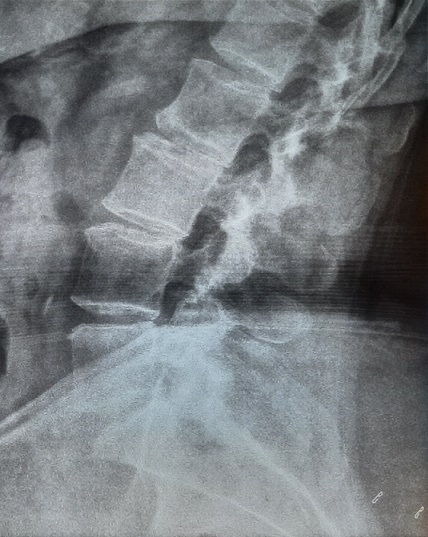
Lumbosacral x-ray
Lumbosacral x-ray. SIDE PROJECTION
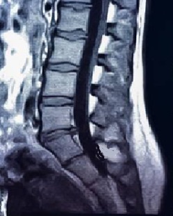
LUMBOSACRAL MRI

RED FLAGS

Red Flags are prognostic variables for serious pathologies. Low back pain due to a serious condition occurs in between 1% and 4% of cases. The diseases involved are: vertebral fracture, neoplasia (1%), infection and cauda equina syndrome.[44]Premkumar, A., et al. “Red Flags for Low Back Pain Are Not Always Really Red: A Prospective Evaluation of the Clinical Utility of Commonly Used Screening Questions for Low Back Pain.” The… Continue reading [45]Greenhalgh, S., and James Selfe. “A qualitative investigation of Red Flags for serious spinal pathology.” Physiotherapy 95.3 2009: 223-226. [46]Verhagen, Arianne P., et al. Red flags presented in current low back pain guidelines. Review. Eur Spine J 25 2016 2788-2802..
If on the one hand the positivity of a red flag can be the indicator of a serious illness, the reverse is not true: the negativity of one or two red flags does not significantly reduce the probability of a diagnosis of serious pathology. The 64% of patients with spinal neoplasms do not have any associated red flags[47]Premkumar, A., et al. “Red Flags for Low Back Pain Are Not Always Really Red: A Prospective Evaluation of the Clinical Utility of Commonly Used Screening Questions for Low Back Pain.” The… Continue reading.
RED FLAGS PER DOLORE LOMBARE
Vertebral fractures[48]Verhagen, Arianne P., et al. “Red flags presented in current low back pain guidelines: a review.” European spine journal 25.9 2016: 2788-2802.
  • History of recent trauma in aged persons >50. The risk is greatest in people aged andevil>70 years
  • use of steroids or immunosuppressants
.
Infections
  • Fever
  • shivering or sweating
  • Recent history of infection
  • Pain that interrupts sleep
  • Persistent night sweats
Cauda equina syndrome
  • Bilateral sciatica
  • Recent loss of control of urinary bladder activity: urinary retention, an impaired flow or impaired awareness of the need to urinate
  • Recent reduction in anal sphincter tone and control
  • Recent loss of bowel control
  • Tingling or numbness in the pubic area and around the anus (saddle anesthesia)
  • Reduction of reflexes in the lower limbs (patellar, achilleo)
Presence of malignant tumor.
It is estimated to occur in less than 1% of patients[49]Henschke, Nicholas, Christopher G. Maher, and Kathryn M. Reef heap. “Screening for malignancy in low back pain patients: a systematic review.” European Spine Journal 16.10 2007: 1673-1679..
  • Previous history of cancer[50]Verhagen, Arianne P., et al. “Red flags presented in current low back pain guidelines: a review.” European spine journal 25.9 2016: 2788-2802.
  • Unintentional weight loss[51]Verhagen, Arianne P., et al. “Red flags presented in current low back pain guidelines: a review.” European spine journal 25.9 2016: 2788-2802.
  • Pain causing nocturnal awakenings
  • Age >50. The risk further increases in patients with age >70

EPIDEMIOLOGY

Most people who experience low back pain that limits activities of daily living experience recurring episodes. Recurrence estimates a 1 year vary from 24% all’80%. The breadth of this range is mainly due to the heterogeneity of the data, for this very reason they must be interpreted with caution[52]Hoy, Damian, et al. “The epidemiology of low back pain.” Best practice & research Clinical rheumatology 24.6 2010: 769-781..

According to a systematic review published in the scientific journal Rheumatology, the prevalence of back pain (regardless of the part of the spine involved) disabling and non-disabling is respectively del 6 he was born in 23% e iThe risk of disabling back pain increases in old age[53]Docking, Rachel E., et al. Epidemiology of back pain in older adults. Prevalence and risk factors for back pain onset. Rheumatology 50 2011 1645-1653..

The onset and course of low back pain is influenced by various environmental and personal factors. L’ the incidence[54]Prevalence: number of cases at a particular instant. Incidence: number of new cases observed in a period of time it is maximum in the third decade of life and prevalence[55]Prevalence: number of cases at a particular instant. Incidence: number of new cases observed in a period of time overall increases with age up to 60-65 years, and then gradually decrease[56]Hoy, Damian, et al. “The epidemiology of low back pain.” Best practice & research Clinical rheumatology 24.6 2010: 769-781..

PHYSIOTHERAPY

In the field of physiotherapy there are several possible methods of intervention, to be adopted according to the individual clinical case:

  • Postural ginnastica Mézières
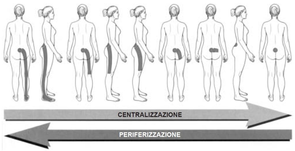
  • Assisted exercises for pain centralization: Centralization is a pain modification achieved with maneuvers that cause peripheral or distal pain to become more centralized (desirable). The opposite (peripheralization of pain) it should neither be sought nor desired[57]Brotzman, S. Brent, and Robert C. Manx. Clinical orthopaedic rehabilitation e-book. An evidence-based approach-expert consult. Elsevier Health Sciences, 2011..
  • Treatment of contractures of the intrinsic spinal muscles
  • Treatment of Myofascial Trigger Points
  • Pumping
  • Tecartherapy (anti-inflammatory and pain reliever)
  • Neuromuscular taping (decontracting, painkiller..)
Da Donelson RG: Mechanical assessment of low back pain. J Musculoskel Med 15[5]:28-39, 1998. Artist: C. Boyter

Bed rest recommendations for the treatment of lower back pain[58]Brotzman, S. Brent, and Robert C. Manx. Clinical orthopaedic rehabilitation e-book. An evidence-based approach-expert consult. Elsevier Health Sciences, 2011.

Royal College of General Practice guidelines[59]To the RCGP 1996 Clinical Guidelines for the Management of Acute Low Back Pain, London, Royal College of General Physicians, 1996.

  • For acute or recurrent DL with or without referred pain in the lower limbs, bed rest for 2-7 days is worse than placebo or ordinary activity. It is not as effective as the alternative treatments it has been compared to for relief from pain, the speed of recovery, the return to daily activities and lost days of work.
  • Prolonged bed rest can cause debilitation, chronic disability and increasing difficulties in rehabilitation.
  • Advice to continue with ordinary activity may result in equivalent or more resolution of acute attack symptoms faster and cause less chronic disability and less time off work than medical treatment “traditional” with pain relievers as needed and advice to rest and let pain guide return to activity normal.
  • A gradual reactivation over days to a few weeks, combined with behavioral pain management, non leads to a big difference in the speed of recovery from pain and disability, but induces less chronic disability e a shorter absence from work.
  • Advice to return to usual work within a short planned time may lead to shorter periods of absence from work.

RECOMMENDATIONS

  • Do not recommend or use bed rest as a treatment for simple back pain.
  • Some patients may be confined to bed for a few days as a result of the pain, but this should not be considered a treatment.
  • Advise patients to remain as active as possible and to continue with normal daily activities.
  • Advise patients to gradually increase physical activities over a few days or weeks.
  • If the patient is working, advising him to stay or return to work as soon as possible is likely to be beneficial.
WORK IN PROGRESS

Note

Note
1 Bagnall, K. M., P. F. Harris, and P. R. Jones. “A radiographic study of the human fetal spine. 1. The development of the secondary cervical curvature.” Journal of anatomy 123.Pt 3 1977: 777.
2 Been, Ella, Sara Shefi, and Michalle Soudack. “Cervical lordosis: the effect of age and gender.” The Spine Journal 17.6 2017: 880-888.
3, 4, 5 Morlachs, A. Mancini, C., and Antonio Mancini. “Orthopedic clinic” Piccin Publisher 1995.
6, 7 Carp, Year, et al. Management of degenerative disk disease and chronic low back pain. Orthopedic Clinics 42.4 2011 513-528.
8, 13, 15, 21, 23, 43 Brotzman, S. Brent, and Robert C. Manx. Clinical orthopaedic rehabilitation e-book. An evidence-based approach-expert consult. Elsevier Health Sciences, 2011.
9, 10, 17, 20 Perolat, Romain, et al. Facet joint syndrome: from diagnosis to interventional management. Insights into imaging 9.5 2018 773-789.
11 Borenstein, David. Does osteoarthritis of the lumbar spine cause chronic low back pain?. Current pain and headache reports 8 2004 512-517.
12 Manchikanti L, Singh V, Pampati V et al 2001 Evaluation of the relative contributions of various structures in chronic low back pain. Pain Physician 4 4 308–316
14 Kalichman, Leonid, et al. Facet joint osteoarthritis and low back pain in the community-based population. Spine 33.23 2008 2560.
16 Perolat, Romain, et al. Facet joint syndrome: from diagnosis to interventional management. Insights into imaging 9.5 2018 773-789.
18 Manchikanti, Laxmaiah, et al. Management of lumbar zygapophysial facet joint pain. World journal of orthopedics 7.5 2016 315.
19 Kalichman L, Kim DH, That L, Germazi A, Hunter DJ 2010 Computed tomography-evaluated features of spinal degeneration. Prevalence, intercorrelation, and association with self-reported low back pain. Spine J 10 3 200–208
22 Brotzman, S. Brent, and Robert C. Manx. Clinical orthopaedic rehabilitation e-book. An evidence-based approach-expert consult. Elsevier Health Sciences, 2011.
24, 32 Hoogendoorn, Wilhelmina E., et al. Systematic Review of Psychosocial Factors at Work and Private Life as Risk Factors for Back Pain. SPINE 25.16 2000 2114-2125.
25 Bernard BP, editor. Musculoskeletal disorders and workplace factors. A critical review of epidemiologic evidence for workrelated musculoskeletal disorders of the neck, upper extremity, and low back. Cincinnati – OH. National Institute for Occupational Safety and Health, US Department of Health and Human Services, 1997.
26 Burdorf A, Sorock G. Positive and negative evidence of risk factors for back disorders. Review. Scand J Work Environ Health 1997;23 4 243-56
27 Fulani, Wilco C., et al. “Influence of gender and other prognostic factors on outcome of sciatica.” Pain 138.1 2008: 180-191.
28, 29, 30, 31 Hoogendoorn, Wilhelmina E. Physical load during work and leisure time as risk factors for back pain. Scand J Work Environ Health 25.5 1999 387-403.
33 Plan, Rahman, et al. “Obesity as a risk factor for sciatica: a meta-analysis.” American journal of epidemiology 179.8 2014: 929-937.
34 Hassan, Ahmed, et al. Chronic low-back pain in adult with diabetes: NHANES 2009–2010. Journal of Diabetes and its Complications 31.1 2017 38-42.
35 Eivazi, Maghsoud, and Laleh Abadi. Low back pain in diabetes mellitus and importance of preventive approach. Health promotion perspectives 2.1 2012 80.
36 Jimenez-Garcia, Rodrigo, et al. Is there an association between diabetes and neck pain and lower back pain? Results of a population-based study. Journal of Pain Research 2018 1005-1015.
37 Huh, Ingrid, et al. Does diabetes influence the probability of experiencing chronic low back pain? A population-based cohort study. The Nord-Trøndelag Health Study. BMJ open 9.9 2019 e031692.
38 Manchikanti, Laxmaiah, et al. Age-related prevalence of facet-joint involvement in chronic neck and low back pain. Pain physician 11.1 2008 67.
39 Schellinger, Dieter, et al. Facet joint disorders and their role in the production of back pain and sciatica. Radiographics 7.5 1987 923-944.
40, 53 Docking, Rachel E., et al. Epidemiology of back pain in older adults. Prevalence and risk factors for back pain onset. Rheumatology 50 2011 1645-1653.
41 Waddell G: The Back Pain Revolution. New York, Churchill Livingstone, 1998.
42 Jensen MC, Brant-Zawadski MN, Obucowski N, et al. Magnetic resonance imaging of the lumbar spine in people without back pain. N Engl J Med. Jul 14; 33 2 69–73, 1994.
44, 47 Premkumar, A., et al. “Red Flags for Low Back Pain Are Not Always Really Red: A Prospective Evaluation of the Clinical Utility of Commonly Used Screening Questions for Low Back Pain.” The Journal of Bone and Joint surgery. American Volume 100.5 2018: 368-374.
45 Greenhalgh, S., and James Selfe. “A qualitative investigation of Red Flags for serious spinal pathology.” Physiotherapy 95.3 2009: 223-226.
46 Verhagen, Arianne P., et al. Red flags presented in current low back pain guidelines. Review. Eur Spine J 25 2016 2788-2802.
48, 50, 51 Verhagen, Arianne P., et al. “Red flags presented in current low back pain guidelines: a review.” European spine journal 25.9 2016: 2788-2802.
49 Henschke, Nicholas, Christopher G. Maher, and Kathryn M. Reef heap. “Screening for malignancy in low back pain patients: a systematic review.” European Spine Journal 16.10 2007: 1673-1679.
52, 56 Hoy, Damian, et al. “The epidemiology of low back pain.” Best practice & research Clinical rheumatology 24.6 2010: 769-781.
54, 55 Prevalence: number of cases at a particular instant. Incidence: number of new cases observed in a period of time
57 Brotzman, S. Brent, and Robert C. Manx. Clinical orthopaedic rehabilitation e-book. An evidence-based approach-expert consult. Elsevier Health Sciences, 2011.
58 Brotzman, S. Brent, and Robert C. Manx. Clinical orthopaedic rehabilitation e-book. An evidence-based approach-expert consult. Elsevier Health Sciences, 2011.
59 To the RCGP 1996 Clinical Guidelines for the Management of Acute Low Back Pain, London, Royal College of General Physicians, 1996.